PATIENT: Norma Valladares Aguilera PHYSICIAN: Dra. Chinchilla CLINIC: Torre Médica Tepeyac BLOCK: KC 16322 IMMUNOHISTOCHEMISTRY: CD3, CD4, CD8, CD20, CD30, CD10, K67 SEX: F AGE: 69 years DATE OF RECEIPT OF MATERIAL: 8/1/2022
DIAGNOSIS:
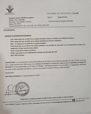
IMMUNOHISTOCHEMISTRY REPORT
* [[File:T cell linfoma.jpg|thumb]]CD3: expressed in about 95% of lymphoid cells including cells with epidermotropism.
* CD4: expressed in about 85% of lymphoid cells of neoplastic appearance.
* CD8: expressed in about 40% of lymphoid cells.
* CD20: expressed in about 0% of lymphoid cells with loss of expression in the component adjacent to the epidermis (i.e., no B-cell marker expression in that region).
* CD30: sporadic positive cells.
* CD10: expressed in a subpopulation of lymphoid cells of approximately 15%.
* Ki67: 80% (high).
COMMENTARY: The immunophenotypic characteristics of the lesion show a marked expression of CD3 and CD4 together with CD7 loss and partial expression of CD8; these findings in the context of the observed histologic features are highly suggestive of a CUTANEOUS T-CELL LYMPHOMA. Immunophenotyping supports a proliferation of T lymphocytes (T primary cells of variable size and pleomorphism), and definitive classification requires correlation with clinical data, morphological study and additional laboratory tests.
DATE: 16/9/2022 MATERIAL RECEIVED: Tissue block KC 16322
Signed and stamped by the pathologist.
(End of translation)
Latest revision as of 17:28, 21 September 2025
Before
Cutaneous T-cell lymphoma recovered with CDS
After
By: Dr. Jorge Ponce – Honduras, Kalcker Institute Master's Student
A 69-year-old female presented with progressive generalized weakness and new-onset cutaneous rashes that prompted initial evaluation at a local health center and subsequent referral to a general hospital for further work-up. Dermatologic assessment and skin biopsy were performed, and immunohistochemical analysis of the lesional tissue confirmed a diagnosis of cutaneous T‑cell lymphoma, establishing a neoplastic T‑cell infiltrate as the cause of her dermatosis. Staging investigations included a chest radiograph that demonstrated cardiomegaly consistent with grade 1 cardiomegaly, an important comorbidity to consider when planning systemic therapies and supportive care. Clinically, the combination of systemic symptoms such as weakness together with chronic or evolving cutaneous lesions should raise suspicion for a cutaneous lymphoma spectrum disorder, and the definitive diagnosis relies on histology supported by immunophenotyping, which in this case proved diagnostic.
Recovered with CDS and DMSO
Enema Protocol
Protocol C-10
Protocol K
REPORT OF PATHOLOGY — TRANSLATION TO ENGLISH
PATIENT: Norma Valladares Aguilera PHYSICIAN: Dra. Chinchilla CLINIC: Torre Médica Tepeyac BLOCK: KC 16322 IMMUNOHISTOCHEMISTRY: CD3, CD4, CD8, CD20, CD30, CD10, K67 SEX: F AGE: 69 years DATE OF RECEIPT OF MATERIAL: 8/1/2022
DIAGNOSIS:
IMMUNOHISTOCHEMISTRY REPORT
CD3: expressed in about 95% of lymphoid cells including cells with epidermotropism.
CD4: expressed in about 85% of lymphoid cells of neoplastic appearance.
CD8: expressed in about 40% of lymphoid cells.
CD20: expressed in about 0% of lymphoid cells with loss of expression in the component adjacent to the epidermis (i.e., no B-cell marker expression in that region).
CD30: sporadic positive cells.
CD10: expressed in a subpopulation of lymphoid cells of approximately 15%.
Ki67: 80% (high).
COMMENTARY: The immunophenotypic characteristics of the lesion show a marked expression of CD3 and CD4 together with CD7 loss and partial expression of CD8; these findings in the context of the observed histologic features are highly suggestive of a CUTANEOUS T-CELL LYMPHOMA. Immunophenotyping supports a proliferation of T lymphocytes (T primary cells of variable size and pleomorphism), and definitive classification requires correlation with clinical data, morphological study and additional laboratory tests.
DATE: 16/9/2022 MATERIAL RECEIVED: Tissue block KC 16322